.jpg)


Understanding the BLUE Protocol in intensive care
Clinical ultrasound plays a crucial role in the case of a patient presenting with a respiratory condition, a common and potentially serious picture that can be caused by a variety of respiratory and cardiovascular pathologies. Using ultrasound, resuscitation physicians can quickly and efficiently assess the lungs, heart and other thoracic structures to identify the underlying cause of symptoms.
Lung ultrasound, for example, can detect abnormalities such as pneumothorax, pleural effusions and pneumonia, while echocardiography can reveal cardiac dysfunctions such as heart failure or valvulopathy. This non-invasive technique enables rapid and accurate management, reducing diagnostic delays and improving the chances of effective treatment and patient recovery.
The BLUE protocol, first described by Daniel Lichtenstein in 2008, quickly became a global benchmark in emergency lung ultrasound. Its simplicity and standardization make it a tool that is suitable for both experienced resuscitators and doctors in training.
In this article, we'll explore the BLUE ultrasound protocol, a valuable tool for intensive care physicians. We'll look at its definition, its decision tree, and its link with PLAPS. We'll also look at the value of the BLUE protocol in the ICU, the different profiles of the protocol, and examples of BLUE examinations.
What is the BLUE protocol in ultrasound?
BLUE (Bedside Lung Ultrasound in Emergency) is a lung ultrasound protocol used in emergency situations. It enables rapid and effective diagnosis of common lung pathologies. The BLUE protocol is based on the analysis of six thoracic points (three bilateral zones) to detect abnormalities such as pneumothorax, pleural effusions, alveolar syndromes (e.g. pneumonia) and interstitial syndromes (e.g. pulmonary edema).
The BLUE protocol is based on a simple principle: "one point, one answer." Systematic analysis of the six chest areas reduces diagnostic uncertainty in more than 90% of cases of acute dyspnea.
Echopneumology is an ultrasound specialty that focuses on lung imaging. The BLUE protocol is a key tool in this specialty, enabling rapid and accurate diagnosis in emergency situations. It is particularly useful for resuscitators, emergency physicians and pulmonologists (who often use an adapted variant of the protocol), who often have to make rapid decisions based on limited information.
The decision tree
The BLUE protocol is based on a decision tree that guides the physician through a series of questions and observations. Here are the key steps in the decision tree:

In practical terms, the decision tree makes it possible to distinguish between respiratory causes (pneumonia, pneumothorax, pulmonary edema) and cardiac causes (left ventricular failure) or mixed causes in just a few minutes. This avoids lengthy and invasive tests and directs the patient straight to the right treatment strategy.
Link with PLAPS Point
The PLAPS point (Postero-Lateral Alveolar and/or Pleural Syndrome) is a specific point used in the BLUE protocol to detect alveolar and pleural abnormalities. It is located on the postero-lateral chest wall and is used to detect pathologies such as pneumonia and pleural effusions. The PLAPS point is a key element in the BLUE protocol decision tree, providing crucial information for diagnosis.
Benefits of the BLUE Protocol in Emergency Ultrasound
The BLUE protocol is particularly useful in intensive care and emergency medicine for several reasons:
- Speed: It enables rapid diagnosis, which is essential in emergency situations.
- Accuracy: It provides precise information on lung pathologies, enabling you to make informed decisions.
- Non-invasive: Ultrasound is a non-invasive technique, making it safe and comfortable for patients.
The BLUE protocol is also particularly useful in resource-limited settings (prehospital care, humanitarian medicine, rural areas). It requires only a portable ultrasound machine and appropriate training, making it an accessible and reproducible method anywhere in the world.
The different BLUE Protocol profiles
The BLUE protocol can be used to diagnose several pulmonary pathology profiles. Here are the main profiles:
- Profile A: Characterized by a normal pleural line and A artifacts. This profile is often associated with pneumothorax.
- B profile: Characterized by B lines, which are vertical artifacts. This profile is often associated with pulmonary edema.
- Profile C: Characterized by alveolar consolidations. This profile is often associated with pneumonia.
- A/B pattern: Characterized by a combination of A and B lines. This pattern is often associated with pneumothorax on the B side.
- PLAPS profile: Characterized by alveolar and pleural abnormalities. This profile is often associated with pneumonia or pleural effusion.
In clinical practice, these profiles are not always exclusive. It is common to observe combinations of signs, particularly in complex cases of acute respiratory distress. Repeating the examination during the course of clinical progression allows the effectiveness of treatment to be monitored and care to be adapted.
Conclusion
The BLUE protocol is an interesting tool for intensive care physicians, emergency physicians and pulmonologists. It enables rapid and accurate diagnosis, using a decision tree based on the analysis of six specific points on the chest wall. The PLAPS point is a key element in this decision tree, providing crucial information for diagnosis. By understanding the different profiles of the BLUE protocol and using concrete examples, healthcare professionals can improve their practice and offer better quality care to their patients.
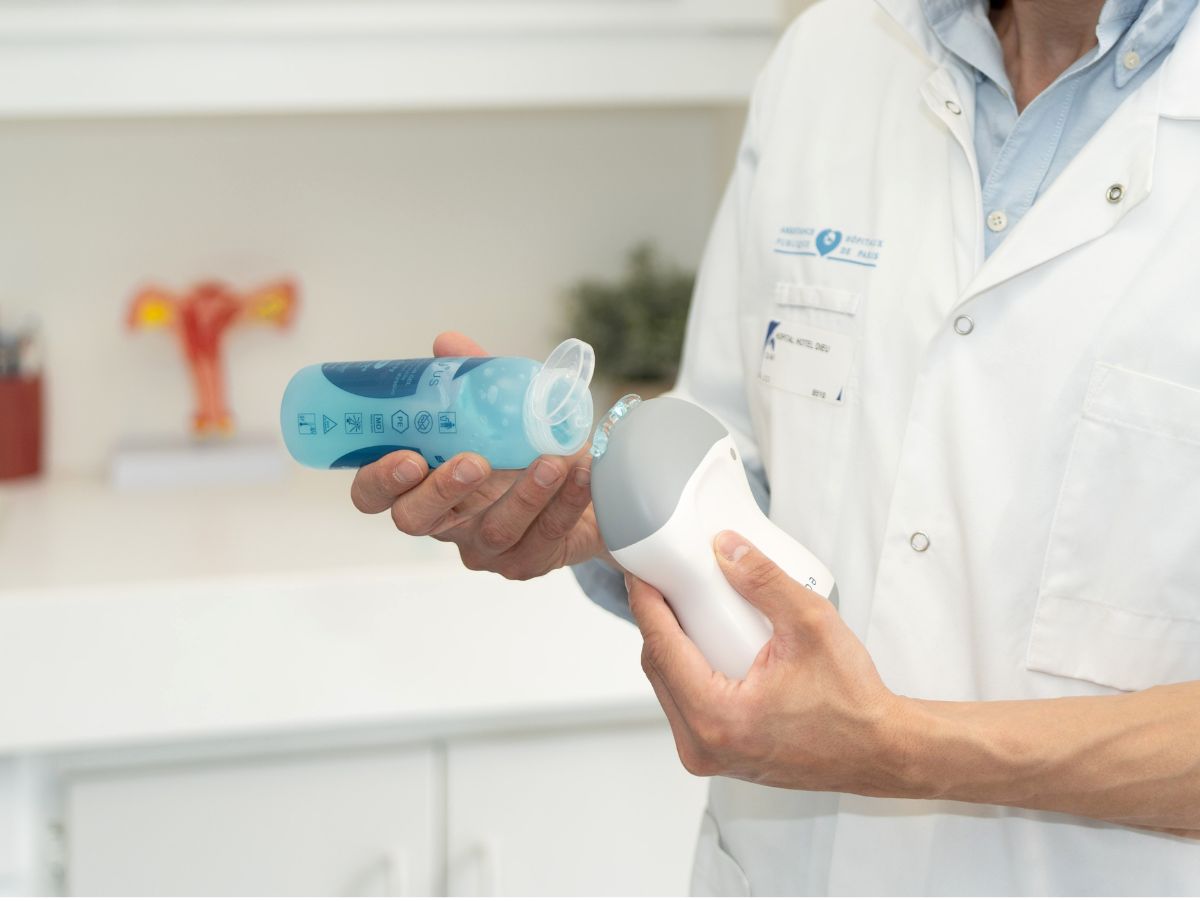
With the widespread adoption of ultra-portable ultrasound devices, such as the echOpen probe, the BLUE protocol is becoming even easier to integrate into daily practice. Lightweight, accessible, and connected, a portable ultrasound device allows the exam to be performed immediately at the patient’s bedside, thereby reinforcing the role of pulmonary ultrasound in modern intensive care.