.png)

10 questions and answers about pleuropulmonary ultrasound
Given the current increase in respiratory infections, the need to streamline imaging tests, and the evolution of the SPLF/SPILF 2025 recommendations, pleuropulmonary ultrasound is emerging as a first-line tool. Accessible, fast, and reproducible, it enables general practitioners to make a more accurate and rapid diagnosis that is better understood by patients. This article answers the 10 most frequently asked questions to support its integration into daily practice.
1. What does the literature say about the role of pleuropulmonary ultrasound in primary care?
Recent literature, notably the 2025 recommendations from the SPLF and SPILF, establishes pleuro-pulmonary ultrasound as a reliable alternative to chest X-rays for diagnosing acute community-acquired pneumonia. It is particularly suitable for primary care, where its speed, lack of radiation, and ability to be performed at the patient's bedside offer a decisive advantage. Reference studies (Orso, Strøm) confirm its excellent diagnostic performance, even when performed by practitioners who are not specialized in imaging.
Beyond pneumonia, ultrasound is also effective in diagnosing pneumothorax and cardiac decompensation. Several meta-analyses (Dahmarde, Ticinesi, Sorensen, Staub) show high sensitivity and specificity, often superior to those of X-rays, with significant time savings in diagnosis.
2. What are the most common use cases?
Three main indications stand out: suspected pneumonia, pneumothorax, and cardiac decompensation with acute dyspnea.
In cases of pneumonia, ultrasound is used to confirm or rule out the diagnosis, particularly in cases where performing a chest X-ray would be time-consuming. In cases of pneumothorax, it allows for rapid and reliable detection, with high specificity that helps to rule out the diagnosis in cases of doubt. Finally, in cases of cardiac decompensation, ultrasound can be used to visualize signs of pulmonary congestion (B-lines, diffuse interstitial syndrome) and quickly guide treatment.
3. How much time and accuracy are saved in diagnosis?
Studies such as that by Nouvenne et al. demonstrate that pleuropulmonary ultrasound significantly improves diagnostic accuracy in cases of acute dyspnea, particularly in elderly patients in long-term care. Nouvenne reports a sensitivity of 94%, a specificity of 90%, and an overall diagnostic accuracy of 92%.
Compared to clinical assessment alone, ultrasound allows for faster identification of the causes of dyspnea: 24 minutes instead of 3 hours, according to a study by Zanobetti et al., with an average examination time of 3-5 minutes according to Amiot et al., whether the cause is pneumonia, cardiac decompensation, or pleural effusion, while reducing the need for radiation-based examinations or biomarkers.
4. Does ultrasound help avoid unnecessary additional tests and antibiotic prescriptions?
A study by Vilar-Palop et al. shows that integrating pleuropulmonary ultrasound into primary care improves the diagnostic accuracy of lower respiratory tract infections while reducing the use of unnecessary additional tests. General practitioners who used ultrasound reported a significant increase in their diagnostic confidence (from 69% to 94%), a reduction in clinical uncertainty, and a change in their treatment decision in nearly 48% of cases. The study also highlights that ultrasound scans made it possible to avoid chest X-rays in approximately 30% of consultations, while reducing the number of unjustified antibiotic prescriptions.
5. What is the patient perception of pleuropulmonary ultrasound in primary care?
The study by Amiot et al. (2024) reveals a high level of confidence and satisfaction among patients who underwent pleuropulmonary ultrasound. Of the 151 patients included, 73.5% were examined by ultrasound, with a median examination time of only 4 minutes. Despite this short format, 81.7% of patients reported a better understanding of their diagnosis, and 82% felt that their general practitioner's medical opinion was more reliable after the ultrasound examination. The stress level associated with the examination remained very low (1.8%), confirming the acceptability of the procedure in an outpatient setting.
6. Can I bill for a pleuropulmonary ultrasound?
Yes, pleuropulmonary ultrasound can be billed under the CCAM nomenclature code GLQP001, which corresponds to a chest ultrasound. This code covers common indications such as pleural effusion, pneumothorax, or pneumonia, provided that the procedure is clinically justified and documented in the medical record.
Billing is possible in addition to the consultation, provided that the ultrasound is a separate procedure performed as part of an independent diagnostic assessment. It is recommended that the clinical factors that led to the examination, the results obtained, and their impact on treatment be specified in the file. This recognition allows physicians to leverage a tool that has become central to respiratory assessment, while complying with regulatory requirements.
7. What is the return on investment for pleuropulmonary ultrasound in general practice?
Pleuro-pulmonary ultrasound is a strategic investment for general medical practices, offering a rapid and measurable return on investment. The cost of a portable ultrasound machine suitable for outpatient practice generally ranges from €1,000 to €10,000, depending on the features. However, each chest ultrasound procedure can be billed in addition to the consultation, with a fee of approximately €30 to €50 per examination, depending on the agreements and the sector of practice.
In practice, a physician performing two to three ultrasounds per day can recoup the cost of their equipment in less than six months, while improving the quality of their care. The study by Amiot et al. (2024) shows that the examination lasts an average of four minutes, which means it can be easily integrated into the consultation flow without significantly extending the medical time.
8. What level of training is required to feel confident in interpreting images?
The study by Amiot et al. shows that 92% of general practitioners trained in pleuro-pulmonary ultrasound feel capable of interpreting images independently in the context of lower respiratory tract infections. After a short, targeted training course, more than 80% consider the tool useful in their daily practice, and nearly 70% say that ultrasound has changed their diagnostic or therapeutic decisions.
9. What training options are available?
The practice of pleuropulmonary ultrasound requires prior training, as specified in the recommendations. The good news is that this training is available to all physicians, even those who are not radiologists. Qualifications vary depending on the specialty and the desired level of competence.
The objective: first acquire the technical skills, then perfect the technique indication by indication. Once you feel confident interpreting the images for a given indication, you will learn how to integrate ultrasound into your clinical reasoning, in addition to the elements of your physical examination.
Available formats:
- Short in-person or video sessions, such as those offered by CFFE, EchoFirst, A2FM, etc.
- E-learning modules with clinical cases (such as those offered by SantéAcadémie)
- Practical workshops at conferences or in healthcare facilities (such as those offered by Winfocus or CHEM)
- On-the-job mentoring with experienced practitioners
- University degrees, to go further (such as those offered by Paris Cité University)
The training courses offered by EchoFirst, Winfocus, CFFE, CHEM, and A2FM are recognized for their quality and tailored to the needs of practitioners. Organized in person or online, they cost an average of €350 for 1 to 2 days, often reimbursable by the DPC. Their content varies according to the level of specialization, with numerous sessions available each year throughout France.
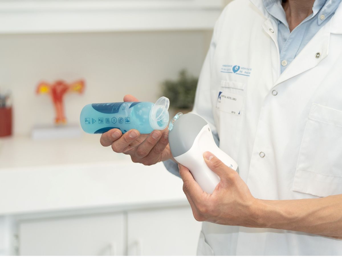
10. Which probe should I buy?
If your goal is to supplement clinical examinations in your office or during visits, the echOpen probe is the ideal solution. Designed for basic ultrasound scans (chest, abdomen, pelvis), it covers the main reasons for consultation without unnecessary complexity. Made in France and supported by the AP-HP, it is designed for general practitioners and emergency physicians who wish to integrate ultrasound into their daily practice without having to obtain a university degree.
With a total cost over five years of €2,976 including tax (probe + subscription), echOpen significantly more affordable than high-end probes (often between €5,000 and €9,500), which offer advanced imaging options to specialized practitioners. It works via an intuitive mobile app, requires only a few hours of training, and allows for rapid image acquisition. Unlike more complex models, it does not overload the practitioner with unnecessary features such as Doppler or 3D, while ensuring robustness and seamless connectivity (Bluetooth/Wi-Fi). With excellent value for money and quick to learn, it is the strategic choice for getting started without compromising on reliability.