.png)

Obstetrical ultrasound training: a key element for midwives
Obstetrical ultrasound has become an indispensable tool in pregnancy monitoring. More and more patients are opting for comprehensive support by midwives, as a complement to, and sometimes, in the case of physiological pregnancies, as an alternative to, follow-up by a gynecologist.
These changes in practice mean that midwives are becoming more skilled, with greater responsibility for prevention, screening and monitoring.
In this context, training in obstetric ultrasound is becoming an essential lever for guaranteeing the quality and safety of care. Thanks to imaging, midwives can objectify several essential conditions and reinforce their autonomy, while respecting their field of competence.
Integrating ultrasound into daily practice is a modern, reassuring response that meets the expectations of both patients and public authorities.
Why is obstetrical ultrasound an essential examination for midwives?
Training in obstetric ultrasound is part of the natural evolution of the midwife's role. For several years now, the profession has benefited from a broadening of its clinical scope, with the possibility of providing comprehensive monitoring of pregnancy, in the context of physiology and early detection of certain anomalies. In this context, ultrasound has become a key skill.
It plays a central role in the pregnancy process: in the first trimester, for confirmation of intrauterine implantation; in the second, for morphological screening; and in the third, for assessment of fetal growth. These stages are essential and form the pillars of modern obstetrical monitoring.
Seeing the fetus, hearing its heartbeat, following its development live: all these elements strengthen the mother-child bond and confidence in the proposed follow-up. For the midwife, ultrasound becomes a clinical decision-making tool, enabling her to anticipate certain complications and refer the patient more quickly if necessary. This ability to act early enhances the quality of follow-up and the safety of care.
How can midwives learn about obstetric ultrasound?
Obstetrical ultrasound: the theoretical basis for midwives
Many organizations now offer training in obstetric ultrasound specifically designed for midwives. These courses are often available as in-service training, either face-to-face or in a hybrid format, and can be adapted to the professional constraints of midwives.
The programs cover all the theoretical fundamentals: reminders of obstetrical anatomy and physiology, physical principles of ultrasound imaging, image reading, identification of fetal structures, growth norms, dating, estimation of amniotic fluid volume, placental localization, and more.
In general, these courses comprise several modules spread over a few weeks to several months, with clearly defined learning objectives: how to perform a dating ultrasound, identify an intra-uterine pregnancy, assess fetal well-being, or recognize signs that may suggest an abnormality and require specialized referral. This rigorous introduction enables midwives to reinforce their expertise in gynecology-obstetrics, and to integrate imaging into their practice in a gradual, supervised manner.
Practice with ultra-portable equipment: the echOpen ultrasound probe
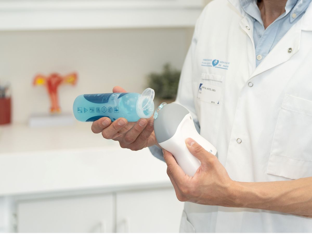
Beyond theory, mastering ultrasound requires regular practice with the right equipment. That’s exactly what the ultra-portable echOpen ultrasound probe offers. Designed for field use, this tri-frequency probe connects wirelessly to a smartphone, fits in a lab coat pocket, and is extremely affordable. It enables midwives to perform certain primary ultrasound examinations—including outside of hospital settings—within the scope of their practice.
In a conventional situation, for example during home pregnancy monitoring or in a birthing center, the midwife can quickly check fetal vitality, the baby's position or the quantity of amniotic fluid, and thus provide additional elements to her clinical examination. With image quality sufficient for rapid clinical assessment, this tool becomes a genuine extension of the physical examination. It facilitates decision-making, secures follow-up and boosts patient confidence.
Common errors that can be avoided with obstetrical ultrasound
Training in obstetrical ultrasound can help you avoid errors with sometimes serious consequences. One of the most frequent errors is pregnancy dating, particularly in the first trimester. However, correct dating determines the rest of the follow-up, from the right timing of examinations to the diagnosis of growth retardation.
Another critical example is the failure to identify an ectopic pregnancy. Thanks to an early ultrasound scan, the trained midwife can suspect abnormal implantation and rapidly refer the patient for specialized medical care, before tubal rupture occurs. Imaging is also essential for detecting a multiple pregnancy, specifying the number of embryos and their type (in terms of choriocity and amnionicity).
Morphological ultrasound, on the other hand, can detect fetal malformations. Although the analysis of these anomalies is the responsibility of the referring physician, the suspicion of atypical signs during a screening examination carried out by a trained midwife can lead to early detection and better management.
A midwife ultrasound scanner can also be used to monitor fetal growth and detect growth anomalies using biometric curves. It plays a key role in assessing amniotic fluid volume, essential for suspecting oligohydramnios or hydramnios.
The tool also enables precise assessment of fetal position, particularly in the case of breech presentation, and locates the placenta to raise the possibility of placenta previa, a frequent source of obstetric complications in the third trimester, and to refer the patient for specialized medical care.