
Lung ultrasound training: a solution for detecting pneumopathy
Pneumonia is a common reason for consulting a general practitioner, particularly among the elderly, immunocompromised patients, and those suffering from chronic conditions. However, conventional diagnostic tools such as standard chest X-rays have many limitations: limited accessibility in primary care, sometimes long waiting times for results, and results that may lack precision or reliability in the early stages. These shortcomings can delay diagnosis, with serious clinical consequences. Unidentified or misdiagnosed pneumonia can lead to rapid deterioration in the patient's condition, avoidable hospitalizations, overuse of antibiotics, and an increased risk of respiratory complications.
In this context, lung ultrasound is emerging as an increasingly recognized tool that can be used as a first-line diagnostic aid for pneumopathies: fast, non-irradiating, and usable directly in the doctor's office or at the patient's bedside, it allows for early detection with good reliability and reproducibility when performed by a professional trained in respiratory pathologies. With appropriate training, healthcare professionals can adopt this tool to enrich their clinical arsenal.
The danger of pneumonias
Pneumonia refers to acute infections of the lung parenchyma. It affects millions of people every year, with a high incidence among at-risk populations: the elderly, immunocompromised patients, and those with chronic diseases (COPD, diabetes, heart failure). In general practice, they are a common reason for prescribing antibiotics and seeking unscheduled medical care.
In terms of public health, community-acquired pneumonia is one of the leading causes of infectious mortality. In France, it is responsible for more than 10,000 deaths annually, mainly among people over the age of 75. This disease burden also results in frequent hospitalizations and increased healthcare costs.
Clinically, pneumopathies vary greatly. While some forms are typical (fever, cough, dyspnea, chest pain), others can be misleading, particularly in elderly or immunocompromised patients, where symptoms are often mild or atypical. This diversity complicates the initial clinical diagnosis and increases the risk of error or delay in treatment. In this context,ultrasound for pulmonologists appears to be a valuable decision-making tool that can be integrated as a first-line tool to guide treatment, in addition to other diagnostic tests.
Diagnosing lung diseases quickly: lung ultrasound
Pulmonary ultrasound is based on simple but powerful principles. It allows the condition of the lung parenchyma and pleura to be analyzed by interpreting ultrasound artifacts. The main ultrasound criteria for pneumonia are subpleural consolidations (hypoechoic, sometimes with air bronchograms), multiple and coalescent B lines (signs of thickened interstitium), and pleural effusion, which is often associated.
Its main advantage lies in its ability to provide a rapid diagnosis, directly at the patient's bedside, without radiation. This makes it a tool that is perfectly suited to general practice, emergency rooms, and outpatient care. Lung ultrasound also allows for dynamic monitoring of clinical progress, with appreciable reproducibility from one examination to the next.
However, like any additional examination, lung ultrasound has its limitations. Good technical skills are essential to interpret the images correctly and avoid false positives. It is therefore intended to complement other examinations (X-ray, CT scan) rather than systematically replace them. Rigorous training in protocols and artifacts is key to its appropriate and safe use.
Training in pulmonary ultrasound: a key issue for tomorrow's practitioners
Training in pulmonary ultrasound is not just a simple introduction: specific training is essential to guarantee the quality of the examination, avoid interpretation errors, and master recognized protocols.Training focuses as much on technical skills (holding the probe, anatomical landmarks) as on semiological interpretation (A lines, B lines, effusions, consolidations).
To meet the needs of general practitioners, specialists, and students, several types of training in chest ultrasound have emerged. Short courses, including intensive practical workshops, enable rapid skill development. E-learning effectively complements theoretical teaching. Finally, tutoring in clinical situations, during internships, or in private practice consolidates what has been learned and facilitates integration into daily practice.
The expected impact of these training courses is considerable: improved detection of pneumonia, reduction in unnecessary hospitalizations, increased skills among primary care physicians, and contribution to more responsive, accurate, and safer medicine. As such, training is a first step, but should not be considered an end in itself: the full integration of lung ultrasound into clinical practice requires a continuous approach.
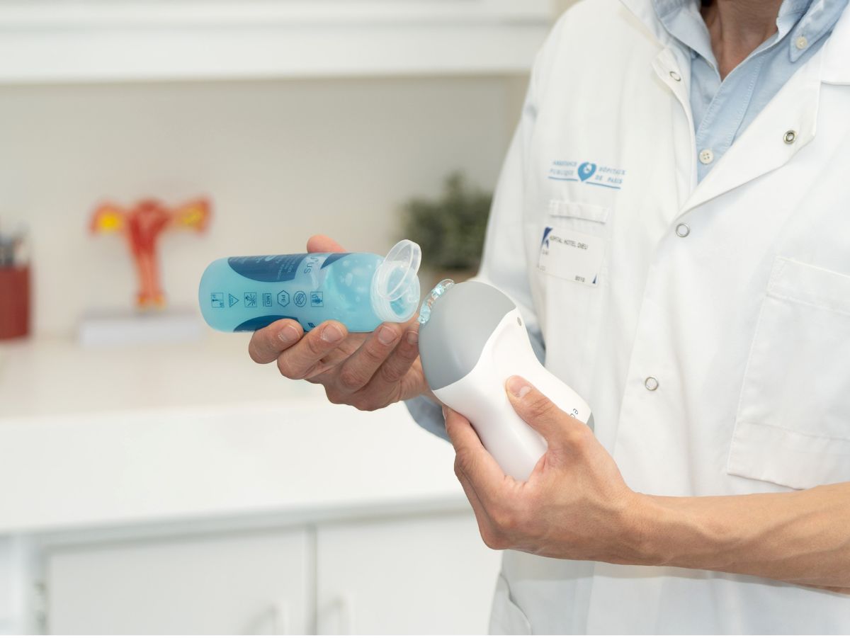
The portable ultrasound device developed by echOpen is part of this trend. Lightweight, intuitive, and connected, it fits in a pocket and allows healthcare professionals to perform initial pulmonary ultrasound exams directly in the office, on the go, or in emergency situations. It is a solution designed to combine performance, accessibility, and continuing education.