Clinical case: prehospital diagnosis of secondary pneumothorax using the echOpen ultraportable ultrasound system
Background
A 73-year-old man presents with a sudden coughing fit, followed by bilateral chest pain resembling tightness. The Mobile Emergency and Resuscitation Service (SMUR) is called by the fire department in response to a desaturation of 85% in ambient air and electrocardiographic abnormalities.
Data collected upon arrival of the SMUR team
- Saturation: 100% at 15 L/min, then reduced to 9 L/min.
- Respiratory rate: 20 cycles/min.
- Heart rate: 120 beats per minute.
- Blood pressure: 120/80 mmHg (normal blood pressure).
- Temperature: apyretic.
- Blood sugar level: euglycemic.
Oxygen therapy is gradually titrated up to 6 L/min.
Electrocardiogram (ECG)
The ECG performed on site shows diffuse ST segment depression, already evident on the reference ECG available. There is a slight increase in V2 to V5, DII, and aVL.
Medical history
- Coronary artery disease with stent in the anterior interventricular artery (AIV), last coronary angiography in January 2025.
- Advanced pulmonary emphysema for five years, following SARS-CoV-2 pneumonia. Ongoing pulmonary follow-up, background treatment withULTIBRO® not taken for three days (stock shortage).
Clinical examination
Cardiovascular system: Regular heart sounds, no murmurs. Peripheral pulses present. No signs of circulatory failure. Calves supple. No edema in lower limbs.
Respiratory system: Absence of vesicular breath sounds on the left; No additional sounds on the right; Respiratory rate of 26 cycles/min; Expiratory braking; Supraclavicular retraction; No cyanosis; No sweating.
Abdomen: Tenderness in the right hypochondrium without mass or guarding.
Neurological: Patient conscious, alert, oriented. Pupils intermediate, symmetrical, reactive.
Performing prehospital lung ultrasound
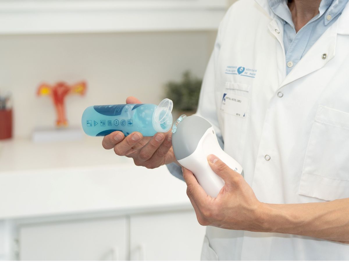
Equipment used: echOpen ultraportable ultrasound probe.
Examination area: left hemithorax.
Results:
- absence of pleural sliding.
- the presence of exclusive A lines (absence of B lines).
Diagnosis: secondary pneumothorax.
Ultrasound videos made with echOpen
Loop 1: absence of pleural sliding in the left hemithorax
Loop 2: line A present, line B absent, reinforcing the hypothesis of pneumothorax
Loop 3: enlarged intercostal space, no visible "lung point"
Hospital confirmation
Upon arrival at the hospital, the diagnosis is confirmed by imaging:
Chest X-ray

Caption: Homogeneous left apical clarity with right mediastinal shift. Appearance consistent with massive left pneumothorax.
Chest scan

Caption: Left compressive pneumothorax with adjacent pulmonary atelectasis. Emphysematous bubbles clearly visible in the right lung parenchyma.
The patient is referred directly for the insertion of a chest drain in a suitable facility.
Contribution of prehospital clinical ultrasound
This case clearly illustrates the benefits of ultra-portable ultrasound in emergency situations:
- Rapid diagnosis, without waiting for complex imaging.
- Direct referral to specialized care.
- Acceleration and optimization of patient care in the emergency room, enabling rapid diagnosis at the patient's bedside without additional delay.
- Decisive therapeutic time savings.
Thanks to the compact, connected, and easy-to-use echOpen device, SMUR responders were able to confirm the diagnosis in the field, which had a direct impact on the patient's care pathway.